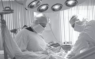
In this practical guide to Lahay Reconstructive Urology, clinicians learn to recognize, prevent, and manage complications that can arise after reconstructive procedures. The goal is to preserve function, minimize patient burden, and support timely recovery. This article outlines key considerations, evidence-based strategies, and stepwise decision-making to help urology teams navigate complications effectively.
Understanding Common Complications in Lahay Reconstructive Urology

Complications in Lahay Reconstructive Urology can include infection, fistula formation, anastomotic leaks, urethral stricture, hematoma, and wound dehiscence. Recognizing patterns associated with specific procedures helps tailor interventions and improve outcomes. A proactive, patient-centered approach reduces delays in treatment and supports healing trajectories.
Key Points
- Early recognition and standardized assessment tools improve outcomes in Lahay Reconstructive Urology complications.
- Multidisciplinary planning, including urology, radiology, wound care, and nursing, reduces time to resolution.
- Tailored reconstructive options depend on tissue availability, prior surgeries, and patient-specific anatomy in Lahay Reconstructive Urology.
- Clear patient communication and shared decision-making enhance adherence to management plans.
- Systematic documentation of complications supports ongoing quality improvement and learning.
Preoperative Planning and Risk Stratification

Identify risk factors such as prior surgeries, radiotherapy, diabetes, smoking, malnutrition, and immune status. Use risk stratification to guide perioperative optimization, antibiotic strategies, and surrogate endpoints for success. In Lahay Reconstructive Urology, preoperative planning should anticipate potential salvage pathways if complications occur.
Intraoperative Techniques to Minimize Complications
Principles include tissue preservation, tension-free anastomosis, meticulous hemostasis, and selective use of interpositions (eg, vascularized flaps). In Lahay Reconstructive Urology contexts, careful flap design and graft choice can influence wound healing and subsequent complication patterns.
Postoperative Management and Early Detection
Adopt standardized drain and catheter protocols, wound care pathways, and early warning signs for prompt escalation. A structured monitoring plan supports timely detection of leaks, infection, or urinary retention and facilitates rapid intervention.
Management Strategies for Specific Complications

Infection and Wound Complications
Prompt antibiotic stewardship guided by culture results, source control for abscesses, and targeted wound care are essential. If infection progresses or pockets form, involve interventional radiology or surgical drainage as needed.
Urethral and Fistulous Complications
Evaluate tissue viability and consider conservative drainage, prolonged catheterization, or staged reconstruction with interposition tissue when indicated by anatomy and prior repairs.
Strictures and Obstruction
Approach may include dilation, endoscopic incision, or revision urethroplasty depending on the location and prior procedures. Ongoing surveillance guides timing of further reconstruction.
Hematoma and Wound Dehiscence
Early hematoma management reduces infection risk. For wound dehiscence, wound optimization, dressings, and selective debridement or negative-pressure therapy may be appropriate in selected cases.
Prosthetic-Related and Urinary Diversion Complications
Monitor for erosion, device malfunction, or diversion-related issues. Plan timely revision or replacement and coordinate with multidisciplinary teams for complex cases.
Clinical Decision-Making Framework
Use a structured framework to guide decisions: assess patient stability, infection status, tissue viability, device function, and patient goals. Plan staged approaches when needed and establish clear timelines for re-evaluation, imaging, and re-intervention.
Postoperative Surveillance and Follow-Up
Schedule follow-ups at 2 weeks, 6 weeks, and 3–6 months, with longer intervals for stable cases. Include imaging or endoscopic evaluation as indicated, and reinforce patient education on warning signs that require prompt reassessment.
What are the most common complications in Lahay Reconstructive Urology?
+Common complications include infection, fistula formation, urinary leakage, urethral or anastomotic strictures, hematoma, and wound dehiscence. Early recognition and multidisciplinary management are key to preserving function and guiding salvage options.
How soon after surgery should a complication be evaluated?
+Red flags such as fever, severe pain, swelling, purulent drainage, or urinary leakage warrant evaluation within 24–72 hours. Subtle signs should prompt assessment within the first two weeks to enable timely intervention and planning.
What imaging or tests are useful in assessing complications?
+Imaging options include ultrasound for collections, CT if infection spreads or an abscess is suspected, voiding cystourethrogram for leaks, urethrography for strictures, and cystoscopy for mucosal assessment. Laboratory tests such as CBC and cultures support diagnosis and guide therapy.
What are first-line management options for a postoperative fistula in Lahay Reconstructive Urology?
+Initial steps include catheter drainage to divert urine, optimized wound care, and infection control. If a fistula persists, plan staged reconstruction with tissue interposition and interval imaging to inform the timing of repair.
How can teams minimize complications in Lahay Reconstructive Urology?
+Focus on preoperative optimization (nutrition, glycemic control, smoking cessation), meticulous surgical technique, appropriate antibiotic stewardship, careful drain management, and structured postoperative surveillance. Regular case reviews foster continuous learning and improvement.